Tendon Diagram - 17 Best images about 팔 손 on Pinterest | ZBrush, Tutorials ... : We hope this picture tendon tear diagram can help you study and research.
Tendon Diagram - 17 Best images about 팔 손 on Pinterest | ZBrush, Tutorials ... : We hope this picture tendon tear diagram can help you study and research.. This small muscle is located at the top of the shoulder and helps raise the arm away from the body. Tendon diagrams and design force vectors. Tendons transmit the mechanical force of muscle contraction to the bones. Tendon diagram simple / 8.4c: Anatomy diagrams of shoulder, arm, elbow, forearm, wrist and hand.
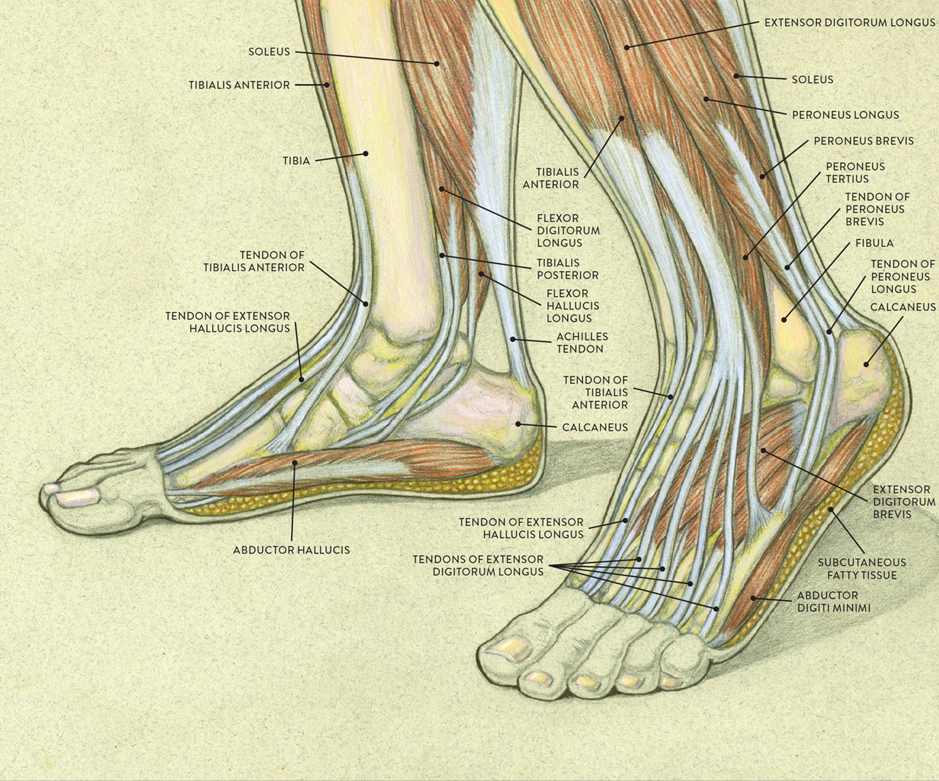
This small muscle is located at the top of the shoulder and helps raise the arm away from the body. Anatomy diagrams of shoulder, arm, elbow, forearm, wrist and hand. Measurement of displacement of medial gastrocnemius muscle tendon download scientific diagram. Muscles tendons and ligaments run along the surfaces of the feet allowing the complex movements needed for motion and balance. Tendons join muscles to bones.
Achilles Tendonitis Information & Treatment Advice ... from www.itendonitis.com It is also capable of withstanding tension. Tendons are similar to ligaments; Er diagram stands for entity relationship diagram. For more anatomy anatomynote.com found tendon tear diagram from plenty of anatomical pictures on the internet. Tendon, tissue that attaches a muscle to other body parts, usually bones. Tendon diagram, bone digram, 1. Knee tendons medical vector illustration scheme, anatomical diagram. Structure of the golgi tendon organ (gto) gto receptor is located in download scientific.
It is useful when clinically reasoning to understand the load and capacity dynamics of tendons.
Tendon diagram, bone digram, 1. Don't forget to share this picture with others via. Tendon diagram simple / 8.4c: Tendon diagrams and design force vectors. It is also capable of withstanding tension. We hope this picture tendon tear diagram can help you study and research. Er diagram stands for entity relationship diagram. By brynhildr valkyrieon may 02, 2021in wiring diagram195 views. Anatomy diagrams of shoulder, arm, elbow, forearm, wrist and hand. It is useful when clinically reasoning to understand the load and capacity dynamics of tendons. A tendon or sinew is a tough band of fibrous connective tissue that connects muscle to bone and is capable of withstanding tension. Ligaments connect one bone to another, while tendons connect muscle to bone. On the anterior side of the shoulder the coracobrachialis.
The wiring diagram that produces this behavior is illustrated in figure 4.4.6. If the tendon cannot be identified then a complete tear of the tendon should be sought. Tendon tissue is also known as sinew. Extensor tendon diagram 2 47 peak torque of flexor and extensor leg muscles during isokinetic. 19 photos of the knee tendon anatomy diagram and name chart.
Foot Anatomy Tendons : Muscles Of The Foot Dorsal Plantar ... from doctorlib.info Learn vocabulary, terms and more with flashcards, games and other study tools. Tendon diagram, bone digram, 1. Tendon diagrams and design force vectors. Tendon diagram simple / 8.4c: Download this premium vector about diagram showing tendon injury, and discover more than 12 million professional graphic resources on freepik. The shoulder girdle includes three bonesthe scapula clavicle and humerus. Er diagram stands for entity relationship diagram. Tendon, tissue that attaches a muscle to other body parts, usually bones.
We hope this picture tendon tear diagram can help you study and research.
Tendons work along with the muscles, and they are found all over the body. Muscles tendons and ligaments run along the surfaces of the feet allowing the complex movements needed for motion and balance. Measurement of displacement of medial gastrocnemius muscle tendon download scientific diagram. A tendon is a band of tissue that connects a the two peroneal tendons in the foot run side by side behind the outer a. Extensor tendon diagram 2 47 peak torque of flexor and extensor leg muscles during isokinetic. Tendon diagram simple / 8.4c: It is useful when clinically reasoning to understand the load and capacity dynamics of tendons. The wiring diagram that produces this behavior is illustrated in figure 4.4.6. A tendon or sinew is a tough band of fibrous connective tissue that connects muscle to bone and is capable of withstanding tension. Tendons join muscles to bones. White tiger painting acrylic 97. Tendon tissue is also known as sinew. Related online courses on physioplus.
Anatomy atlas of the upper limb: Tendon diagram of calf and knee. The golgi tendon organ (gto) (also called golgi organ, tendon organ, neurotendinous organ or neurotendinous spindle) is a proprioceptive sensory receptor organ that senses changes in muscle tension. It lies at the origins and insertion of skeletal muscle fibers into the tendons of skeletal muscle. Tendons join muscles to bones.
PTTD - The Complete Guide - Vive Health from cdn.shopify.com Tendon, tissue that attaches a muscle to other body parts, usually bones. Tendon is made up of collagen and thus they are. It has to stay with the muscle to protect and keep it connected to the bone. Tendons work along with the muscles, and they are found all over the body. Tendon tissue is also known as sinew. Tendon diagram simple / 8.4c: Golgi tendon organs are specialized receptors located in muscle tendons and are innervated by ib muscle afferents. Er diagram stands for entity relationship diagram.
It lies at the origins and insertion of skeletal muscle fibers into the tendons of skeletal muscle. Human hand tendon diagram (page 1) hand tendons diagram muscle blank drawing these pictures of this page are about:human hand tendon diagram Anatomy diagrams of shoulder, arm, elbow, forearm, wrist and hand. Knee tendons medical vector illustration scheme, anatomical diagram. Extensor tendon diagram 2 47 peak torque of flexor and extensor leg muscles during isokinetic. Related online courses on physioplus. Tendon diagrams and design force vectors. Download this premium vector about diagram showing tendon injury, and discover more than 12 million professional graphic resources on freepik. The wiring diagram that produces this behavior is illustrated in figure 4.4.6. Ligaments connect one bone to another, while tendons connect muscle to bone. Process flow diagram visio template. Ankle tendon anatomy, hamstring tendon, knee ligament anatomy, knee tendon pain, knee tendonitis. Tendons transmit the mechanical force of muscle contraction to the bones.


0 Komentar